Which of these images shows hyaline cartilage? With this intriguing question at the forefront, we embark on an enlightening journey into the realm of cartilage, exploring its unique characteristics, diverse locations, and pivotal role in joint function. Join us as we delve into the fascinating world of hyaline cartilage, uncovering its intricate structure, remarkable properties, and clinical significance.
Hyaline cartilage, a specialized connective tissue, stands out with its exceptional resilience and smooth, glassy appearance. Its unique composition grants it the ability to withstand compressive forces, making it an essential component of various structures throughout the body, including the articular surfaces of joints, the rib cage, and the delicate framework of the nose.
Its remarkable properties contribute to the smooth, effortless movement of joints, the protection of delicate tissues, and the overall structural integrity of the body.
Characteristics of Hyaline Cartilage

Hyaline cartilage is a type of cartilage that is characterized by its smooth, glassy appearance. It is the most common type of cartilage in the body and is found in many different locations, including the joints, the ribs, and the nose.
Hyaline cartilage is composed of chondrocytes, which are cells that produce and maintain the cartilage matrix. The matrix is made up of collagen fibers and proteoglycans, which give hyaline cartilage its strength and flexibility.
Hyaline cartilage differs from other types of cartilage in several ways. First, it is the only type of cartilage that is avascular, meaning that it does not contain any blood vessels. This means that hyaline cartilage must rely on diffusion for the exchange of nutrients and waste products.
Second, hyaline cartilage is the most flexible type of cartilage. This flexibility is due to the high content of proteoglycans in the matrix.
| Characteristic | Description |
|---|---|
| Appearance | Smooth, glassy |
| Location | Joints, ribs, nose, and other locations |
| Cell type | Chondrocytes |
| Matrix composition | Collagen fibers and proteoglycans |
| Vascularity | Avascular |
| Flexibility | Most flexible type of cartilage |
Locations of Hyaline Cartilage

Hyaline cartilage is found in many different locations in the body, including:
- Joints:Hyaline cartilage covers the ends of bones in joints. It helps to reduce friction and wear and tear on the bones.
- Ribs:Hyaline cartilage connects the ribs to the sternum. It helps to protect the chest cavity and the organs within it.
- Nose:Hyaline cartilage forms the shape of the nose.
- Trachea:Hyaline cartilage helps to keep the trachea open.
- Larynx:Hyaline cartilage forms the vocal cords.
Hyaline cartilage is also found in some other locations, such as the ears and the intervertebral discs.
| Location | Function |
|---|---|
| Joints | Reduces friction and wear and tear on bones |
| Ribs | Connects ribs to sternum and protects chest cavity |
| Nose | Forms shape of nose |
| Trachea | Keeps trachea open |
| Larynx | Forms vocal cords |
Role of Hyaline Cartilage in Joint Function

Hyaline cartilage plays an important role in joint function. It helps to:
- Reduce friction:The smooth surface of hyaline cartilage helps to reduce friction between the bones in a joint. This helps to prevent wear and tear on the bones and allows for smooth movement.
- Absorb shock:Hyaline cartilage is also able to absorb shock. This helps to protect the bones and joints from damage.
- Load bearing:Hyaline cartilage helps to bear the load of the body. This helps to prevent the bones from collapsing under pressure.
Hyaline cartilage is an important part of the joint system. It helps to keep the joints healthy and functioning properly.
Illustration:The diagram below shows the role of hyaline cartilage in joint function.
[Diagram of a joint showing the hyaline cartilage]
Histological Analysis of Hyaline Cartilage: Which Of These Images Shows Hyaline Cartilage

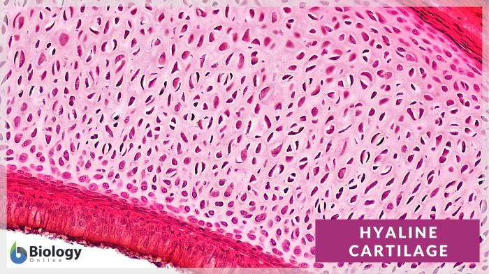
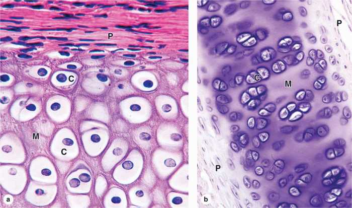
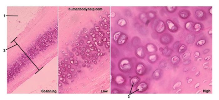
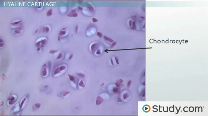
Hyaline cartilage can be distinguished from other types of cartilage by its histological appearance. Under a microscope, hyaline cartilage appears as a clear, glassy substance with no visible cells. This is due to the fact that the chondrocytes are located in small cavities within the matrix.
The matrix is composed of collagen fibers and proteoglycans, which give hyaline cartilage its strength and flexibility.
The chondrocytes in hyaline cartilage are responsible for producing and maintaining the matrix. They are also responsible for regulating the exchange of nutrients and waste products between the cartilage and the surrounding tissues.
Photomicrograph:The photomicrograph below shows a histological section of hyaline cartilage.
[Photomicrograph of hyaline cartilage]
Clinical Significance of Hyaline Cartilage
Hyaline cartilage is an important tissue in the body. It plays a vital role in joint function and helps to protect the bones from damage. However, hyaline cartilage can be damaged by a variety of factors, including:
- Trauma:Hyaline cartilage can be damaged by trauma, such as a fall or a blow to the joint.
- Osteoarthritis:Osteoarthritis is a degenerative joint disease that can damage hyaline cartilage. It is the most common type of arthritis.
- Rheumatoid arthritis:Rheumatoid arthritis is an autoimmune disease that can damage hyaline cartilage. It is less common than osteoarthritis.
Damage to hyaline cartilage can lead to joint pain, stiffness, and swelling. It can also lead to decreased range of motion and difficulty walking. In severe cases, damage to hyaline cartilage can lead to joint replacement surgery.
There are a number of treatments available for hyaline cartilage damage. These treatments include:
- Medication:Medication can be used to relieve pain and inflammation.
- Physical therapy:Physical therapy can help to improve range of motion and strength.
- Surgery:Surgery may be necessary to repair or replace damaged hyaline cartilage.
The prognosis for hyaline cartilage damage depends on the severity of the damage. With early diagnosis and treatment, it is possible to prevent further damage and maintain joint function.
FAQ Summary
What is the main function of hyaline cartilage?
Hyaline cartilage plays a crucial role in joint function, providing a smooth, gliding surface for joint movement, absorbing shock, and distributing weight-bearing forces.
Where is hyaline cartilage found in the body?
Hyaline cartilage is found in various locations throughout the body, including the articular surfaces of joints, the rib cage, the nose, and the trachea.
How does hyaline cartilage differ from other types of cartilage?
Hyaline cartilage is distinguished from other types of cartilage by its smooth, glassy appearance, its lack of blood vessels and nerves, and its high content of collagen fibers.